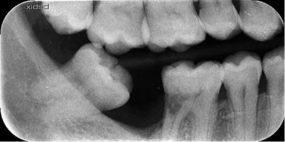
It is a conservative procedure to remove pulp tissue (the nerve of the tooth) affected by decay, infection or trauma. After thorough disinfection, the canals are properly sealed, preserving the chewing function and appearance of the tooth.
The challenge is working in a small space with difficult access; that is why we use the microscope, which also helps to locate narrow canals and to see complex anatomy. This allows us to be more conservative and to work with maximum precision, as the microscope magnifies the image up to 26 times compared with normal vision.
The process, step by step
- 01
First visit
We assess your case, listen to what concerns you and explain which tests we need.
- 02
Diagnosis and plan
We study the information and present you with the plan, the timescales and the quote.
- 03
Treatment
We carry it out over the agreed sessions.
- 04
Maintenance
We schedule check-ups to make sure the result remains stable over time.